The thermal paradox the slide is built around
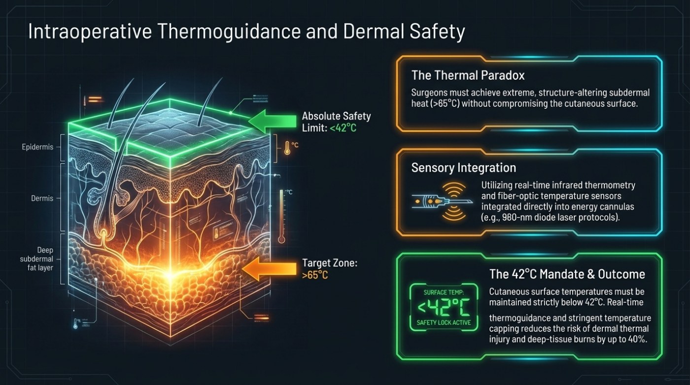
The seventh slide opens with a contradiction stated as plainly as the deck can state it: surgeons must achieve extreme, structure-altering subdermal heat above 65 °C without compromising the cutaneous surface. Those two requirements pull in opposite directions. The layer that has to be hot is a few millimetres away from the layer that must stay cool, and the tissue between them conducts. Everything else on the slide — the layer diagram, the sensors, the capped surface temperature — exists to manage that distance. If you have been weighing up fat removal surgery for a home around Sarasota, FL, this guide covers how it actually works, what it tends to cost, and where the results usually fall short.
The short version
- The slide frames a paradox: surgeons must reach subdermal heat above 65 °C without letting the cutaneous surface exceed 42 °C.
- The two figures describe a window rather than a setting, because the layers between them conduct heat.
- Real-time infrared thermometry measures the surface without contact, and fibre-optic sensors sit inside the energy cannulas.
- A surface display reading below 42 °C with a safety lock active turns a measurement into a stop condition.
- The deck attaches a risk reduction of up to 40 percent for dermal thermal injury and deep-tissue burns to strict capping.
The paradox is worth restating in patient terms. A technique that claims to tighten the skin by heating tissue is also, by definition, a technique that puts heat close to the skin. What separates a controlled thermal effect from a burn is not the temperature at the tip of the device; it is whether the surface temperature is being watched while the heat is delivered, and whether the delivery stops when the surface reaches its limit.
The layer-by-layer temperature map
The slide's diagram is a cross-section labelled in three depths: the epidermis at the surface, the dermis beneath it, and the deep subdermal fat layer at the bottom. Two temperatures are pinned to those layers. A green arrow pointing at the surface is labelled Absolute Safety Limit: <42 °C. An orange arrow pointing into the deep subdermal fat layer is labelled Target Zone: >65 °C. The epidermal and dermal layers sit in the gradient between them.
| Layer in the diagram | Temperature shown | How the slide labels it |
|---|---|---|
| Epidermis, at the surface | Below 42 °C | Absolute safety limit, with the surface reading displayed as a locked value |
| Dermis, beneath the surface | Between the two extremes | Shown as the transition zone the gradient crosses |
| Deep subdermal fat layer | Above 65 °C | Target zone — the layer the energy is aimed at |
| The distance between them | Not given in millimetres | Presented as the reason monitoring is required rather than assumed |
Read as a pair, the two figures describe a window rather than a setting. More than 65 °C has to be reached at depth, and less than 42 °C has to hold at the surface, at the same time, in adjacent tissue. That is the specification the rest of the deck's safety content is measured against, and our published guide on heat-based tightening describes the same two numbers as numbers that only make sense together, because everything between them is the safety problem.
Sensory integration: how the numbers are supposed to be known
A temperature target is meaningless unless it is being measured during the procedure, and the slide's middle panel says how: real-time infrared thermometry and fibre-optic temperature sensors integrated directly into the energy cannulas, with 980 nm diode laser protocols named as the example. The distinction being drawn is between measuring the device and measuring the tissue, and the slide puts the sensor in the instrument that is inside the patient.
| Element | As described on the slide | What it is there to catch |
|---|---|---|
| Real-time infrared thermometry | Non-contact measurement of surface temperature during delivery | A surface that is climbing towards the 42 °C limit while energy is still being applied |
| Fibre-optic temperature sensors | Integrated directly into the energy cannulas | Temperature at the working depth rather than at the device's outer housing |
| 980 nm diode laser protocols | Named on the slide as the example platform for this sensor arrangement | Dosing decisions that depend on a reading taken at the point of delivery |
| Surface temperature display | Shown on the slide reading below 42 °C with a safety lock active | An operator continuing past a limit because nothing told them to stop |
The last row is the one that turns monitoring into safety. A number on a screen is information; a lock that acts on the number is a control. The slide presents the display as an interlock with the word active next to it, which is the difference between watching a value and being governed by it.
The 42 °C mandate and the 40% figure
The bottom panel of the slide states the rule and its intended consequence. Cutaneous surface temperatures must be maintained strictly below 42 °C. Real-time thermoguidance and stringent temperature capping, the slide continues, reduces the risk of dermal thermal injury and deep-tissue burns by up to 40 %. The word cap is doing the work again: the surface temperature is being held under a ceiling rather than watched until something goes wrong.
| Claim on the slide | Exactly as stated | What a reader should not infer |
|---|---|---|
| The surface rule | Cutaneous surface temperatures maintained strictly below 42 °C | That a single reading below the limit at any one moment is sufficient |
| The mechanism of control | Real-time thermoguidance and stringent temperature capping | That monitoring without a stop rule produces the same risk reduction |
| The risk reduction | Up to 40 % for dermal thermal injury and deep-tissue burns | That the reduction is measured, or that it applies to every platform and every patient |
| Source of the figure | Presented as the deck's own summary of the technique | That a named study with a sample size and a comparison group sits behind the percentage |
The percentage deserves the same treatment as the temperatures. It is a figure from the source material behind this deck, not an independently reproduced trial result, and our published guide notes the same limitation for the thermal numbers generally. What can be said without overreach is directional: the slide's position is that continuous measurement plus a hard cap is safer than intermittent attention, and that the difference is the point of the protocol rather than an optional extra. The deck does not break the 40 % down by injury type, and this article will not either.
Why the surface rule is the hard one
The subdermal target is a goal; the surface limit is a constraint. Those behave differently in practice. A goal can be approached gradually and confirmed when it is met. A constraint has to hold for the entire procedure, including the moments when the operator is concentrating on contour rather than on the monitor, which is why the slide's language moves from measurement to an interlock.
It is also the reason this page sits immediately after the slide on thermal contraction and immediately before the slide on operating-room controls. The thermal effect is the objective, the surface cap is the boundary condition, and the room the procedure happens in is the environment the heat balance has to work in. The deck treats all three as one protocol rather than three topics.
Questions this slide raises
Why is the surface limit so far below the subdermal target?
The slide sets the two figures for different tissue. More than 65 °C is described as the level at which the subdermal collagen response begins. Below 42 °C is described as the absolute surface limit — not a working temperature but a ceiling, and the design of the whole monitoring arrangement follows from the size of the gap between a target and a limit.
What does temperature capping actually change?
It converts a measurement into a stop condition. The slide shows both: real-time thermometry and fibre-optic sensors producing the reading, and a surface display with a safety lock active holding the value below 42 °C. Without the second half, the first is a number printed on a monitor.
Does the 40 % figure apply to every technique?
The slide states it as the reduction associated with real-time thermoguidance and stringent temperature capping, describing dermal thermal injury and deep-tissue burns. It is not presented with a study name, a sample size or a platform comparison, so it is best read as the source material's own summary of why the protocol exists rather than as a rate that transfers to any particular device setting.
This is published information, not medical advice — a board-certified surgeon must assess whether a procedure suits you.
Related Guides
Three guides that pick up where this slide stops:
- Heat-Based Skin Tightening After Liposuction: What the Numbers Mean — the same 65 °C target and 42 °C surface limit, and what they do and do not prove.
- Liposuction Risks and Complication Rates as Published — how published complication figures are counted, and why they differ between sources.
- Verifying Surgeon Credentials and Facility Accreditation — what to check about who is operating and where the procedure is being performed.